There is considerable interest in developing optical microscopes presenting a lateral resolution below the usual Rayleigh criterion, λ/(2NA), where λ is the wavelength of the illumination and NA is the numerical aperture of the imaging system, while retaining the convenience of far-field illumination and collection. Among the various ways to ameliorate the resolution, it has been proposed to illuminate the sample with many structured illuminations, namely standing waves, and to mix the different images through simple arithmetics.This technique is very close to optical diffraction tomography (ODT) in which the sample is illuminated under various angles of incidence, the phase and intensity of the diffracted far-field is detected along several directions of observation, and a numerical procedure is used to retrieve the map of the permittivity distribution of the object from the far-field data. Experimental and theoretical studies have shown that using several illuminations permits one to exceed the classical diffraction limit by a factor of two.
In all microscopy techniques using several successive illuminations, one needs a numerical procedure to combine the different images and extract the map of the relative permittivity distribution of the object from the scattered far-field. In general, one assumes that the object is a weak scatterer so that there is a linear relationship between the scattered field and the relative permittivity of the object, namely one assumes that the Born approximation is valid. In this case, the transverse resolution limit can be inferred from simple considerations on the portion of the Ewald sphere that is covered by the experiment. It is limited by λ/2(ni+nd) for configurations in which the incident waves propagate in a medium of refractive index ni, while the diffracted waves propagate in a medium of refraction index nd.
In this web page we present a brief overview of our research in optical diffraction tomography. For more details on each topic have a look at the references.
Superresolution in total-internal reflection tomography
Influence of multiple scattering with optical diffraction tomography
Beyond the Rayleigh criterion : Grating assisted far-field optical diffraction tomography
Experimental Demonstration of Quantitative Imaging beyond Abbe's Limit
Mirror-assisted optical diffraction tomography
Nanometric resolution using far-field optical tomographic microscopy
Tomographic diffractive microscopy with a wavefront sensor
Tomographic diffractive microscopy achieves a resolution about λ/4
Superresolution in total-internal reflection tomography
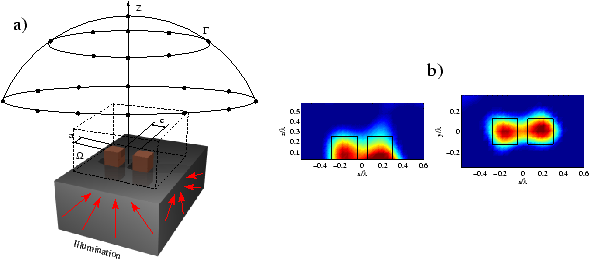
We simulate a three-dimensional optical diffraction tomography experiment in which superresolution is achieved by illuminating the object with evanescent waves generated by a prism and the amplitude and phase of the scattered fields are detected above the subtrate (see figure below on the left side). We show the superresolution is attained by illuminating the sample with evanescent waves and taking into account the multiple scattering between the objects and the interface in the non linear inversion procedure. Figure below on the left side show reconstructed relative permittivity distribution with the nonlinear inverse algorithm from corrupted data with noise when evanescent waves are used as incident waves. We clearly retrieved the two cubes separated by a distance center to center about λ/5.

References :
Influence of multiple scattering with optical diffraction tomography
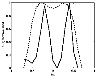
Most reconstruction procedures in optical diffraction tomography assume that single scattering is dominant so that the scattered far field is linearly linked to the permittivity. The nonlinear inversion that we used (based on the rigourous solution of the Maxwell’s equations), is applied to complex three-dimensional samples. We have shown that multiple scattering permits one to obtain a power of resolution beyond the classical limit imposed by the use of propagative incident and diffracted waves. The illumination and the scattered field are plane waves propagating toward positive z (transmission configuration). The figure below shows the normalized reconstructed permittivity- along the z axis for two dipoles (no multiple scattering) and two cubes of width λ/4 with high permittivity equal to 2.25. The sepration center to center is equal to 0.6λ. We observe that the two dipoles are not resolved (dashed line) while the cubes are easily distinguished (solid line). In our opinion, the presence of multiple scattering and the use of a nonlinear inversion scheme is responsible for the better resolution of the image of the two cubes.
Reference :
Beyond the Rayleigh criterion : Grating assisted far-field optical diffraction tomography
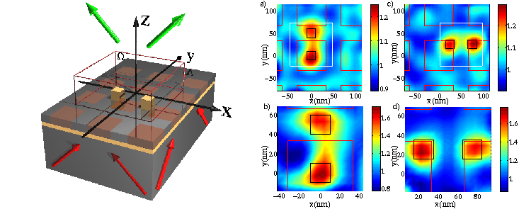
We propose an optical imaging system, in which both illumination and collection are done in far field, that presents a power of resolution better than one-tenth of the wavelength. This is achieved by depositing the sample on a periodically nanostructured substrate illuminated under various angles of incidence as showed in the figure below on the right side (successively illuminated from below and the far field is detected above the substrate). The superresolution is due to the high spatial frequencies of the field illuminating the sample and to the use of an inversion algorithm for reconstructing the map of relative permittivity from the diffracted far field. Thus, we are able to obtain wide-field images with near-field resolution without scanning a probe in the vicinity of the sample. Fgures below on the rigth side show the map in the (x, y) plane of the permittivity for different positions of the objects with respect to the grating. We can see that the two cubes separated by a distance center to center of λ/10 are correctly retrieved.
References :
Experimental Demonstration of Quantitative Imaging beyond Abbe's Limit
We show experimentally that the optical diffraction tomography permits us to obtain the map of permittivity of highly scattering samples with axial and transverse resolutions that are much better than that of a microscope with the same numerical aperture. The sample is constituted of two rectangular rods of resin deposited on a silicon substrate. The rods height is 110 nm and width 200 nm separated by 300 nm side to side. If we have a look at the figure on the rigth we have : (a) Height profile provided by the AFM. (b) Dotted line : squared modulus of the reflectance obtained with ODT-LIA approach. Solid line : Intensity measured at the image plane of a wide-field optical microscope with NA=0,75 and red incoherent light. (c) Map of the permittivity obtained with the Non-Linear Inversion Algorithm applied to the same data as that used in the ODTLIA approach. (d) Comparison along the dashed line plotted in (c) of the reconstructed permittivity (dashed line) with the actual value (solid line). Note that the separation between the rods is so marked that it is likely that 400 nm is not the ultimate resolution of the imager. In our opinion, the significant improvement brought by our method is essentially due to the fact that our method is based on a rigorous calculation of the diffracted field and it has potentially access to spatial frequencies that are higher than that given by the single-scattering analysis.

Reference :
Mirror-assisted optical diffraction tomography
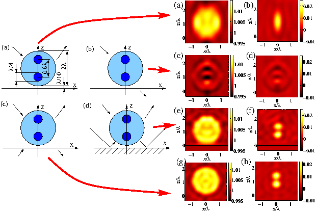
We demonstrate that the axial resolution of a reflection tomographic diffractive microscope is drastically improved when the sample is placed in front of a perfect mirror. We show analytically and with rigorous simulations that this approach permits us to obtain images with the same isotropic resolution as that obtained when the sample is illuminated and observed from every possible angle. Figure below on the left side shows the four different configuration under study (a : reflection b : transmision c : isotropic d : mirror) and figure below on the right side the reconstruction obtained real and imaginary part of the relative permittivity. The link between the configuration and its reconstruction is done by a red arrow. This clearly show that the presence of a mirror permits us to get an isotropic resolution like with a complete configuration : the last two lines in the left figure are similar.
Reference :
Nanometric resolution using far-field optical tomographic microscopy
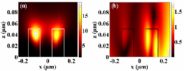
The resolution of optical far-field microscopes is classically diffraction-limited to half the illumination wavelength. We show experimentally that this fundamental limit does not apply in the multiple scattering regime. We used tomographic diffractive microscopy to image two pairs of closely spaced rods (with a width and interdistance of 50 nm=λ/12) of widely different diffractive properties. We use an inversion algorithm accounting for multiple scattering.The figure below shows (left figure) the reconstruction obtained from experimental data of two germanium rods spaced by 100 nm, with width of 100 nm and height of 50 nm with a wavelength of illumination of 632.8 nm (the white line represents the actual height profile). The simulated total field modulus in the figure (right side) shows that multiple scattering yields subdiffraction bright spots that are likely to probe the fine details of the object. with low relative permittivity and high relative permittivity. This achievement demonstrates that the power of resolution of digital far-field microscopes in the multiple scattering regime can be much better than the classical Abbe limit provided inversion algorithms based on a rigorous modeling of the wave-sample interaction are used.
Reference :
Tomographic diffractive microscopy with a wavefront sensor
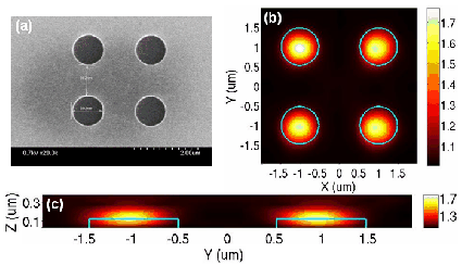
We show that, using a wavefront sensor, tomographic diffractive microscopy can be implemented easily on a conventional microscope, as it is not necessary in that case, to add a reference beam to the setup and recording an interference pattern in an on-axis or an off-axis arrangement. Moreover, the number of illuminations can be dramatically decreased if a constrained reconstruction algorithm is used to recover the sample map of permittivity. In the figure below we compare the reconstruction obtained with (a) Electron microscope image of the sample ; and our non linear algorithm from data obtained with a wavefront sensor : (b) Transverse permittiv ity cut at a height of 125 nm of the reconstruction ; (c) Longitudinal permittivity cut.
Reference :
Tomographic diffractive microscopy achieves a resolution about λ /4
We have developed the first optical digital microscope that exploits all the information accessible via the diffraction process : intensity, phase, and polarization state of the scattered field for any possible illumination within the numerical aperture of the objective. In the single scattering regime, this ultimate microscope is able to reconstruct permittivity maps with a resolution about one-fourth of the wavelength. This experimental achievement, which outperforms that of all existing far-field microscopes, points out the importance of accounting for light polarization when tackling super resolution and sets a landmark in optical far-field imaging.
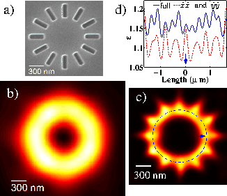
To document further the potential of the full-polarized TDM, we image a complex sample made of twelve resin rods of width 100 nm, length 300 nm, and height 140 nm radially placed at the summit of a dodecagon [Fig. 7(a)]. The sample is illuminated by eight directions of incidence, defined by a fixed polar angle 60- degres and an azimuthal angle regularly spaced within 360 degres. (a) Scanning electron microscope image. (b) Dark-field optical microscope image. (c) Reconstructed permittivity averaged over the sample’s height using full-polarized TDM data. (d) Permittivity along the dashed circle in (c). Plain line : full-polarized data ; dashed line : the combined scalar data.
Reference :