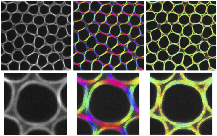
Abstract : Genetically engineered tools help scientists watch how actin filaments arrange inside living cells and tissues. Animal life relies on a protein called actin. Actin helps cells and tissues stay intact and hold their shape. It plays an important role in several processes that are crucial for animals to grow and function. For example, actin helps cells divide and move, and muscles contract. Actin is able to perform all these tasks because it can form long, thread-like structures, called actin filaments, that organize in a specific way to accomplish the different functions. Biologists typically visualize actin filaments in living cells by using fluorescent proteins. Fluorescent proteins are special proteins that can absorb light of one color and then emit light of a different color. The most famous one is GFP (Green Fluorescent Protein, awarded a Nobel Prize in 2008), which glows green when exposed to blue light. By attaching these fluorescent proteins to actin inside a cell, researchers are able to watch actin filaments in action in real time using a technique called fluorescence microscopy. While this approach is very powerful for showing where filaments are located, it falls short of revealing how they are arranged.

Fluorescence polarization microscopy (or polarimetry) is an advanced microscopy method developed by optical physicists at Institut Fresnel, which is uniquely placed to measure how actin filaments are arranged inside cells. However, for polarimetry to work, the glowing protein (GFP) attached to actin needs to stay mostly still—unfortunately, the GFP is too flexible, making it unsuitable for this technique. Biologists at Institut Fresnel teamed up with biochemists, structural and cell biologists and biophysicists in France (IBPS, IBDM, IJM, IPBS, CRCT) and abroad (University of Toronto and IBFG/University of Salamanca) to take on the challenge to immobilize the GFP. This teamwork succeeded to constrain the mobility of GFP, creating adapted genetically engineered tools that can be used with polarimetry to watch how actin filaments arrange and rearrange in real time inside living cells and tissues to accomplish their functions. These tools, along with the freely shareable (open source) data analysis software, PyPOLAR, co-developed with the Mathematics Institute of Marseille (I2M), make a powerful set of resources that promise to improve our understanding about how actin filaments help animals live and function.
Article in French on CNRS Ingénierie website : https://www.insis.cnrs.fr/fr/cnrsinfo/comment-les-filaments-dactine-sorganisent-linterieur-des-cellules-et-des-tissus-vivants
Reference : Carla Silva Martins, François Iv, Shashi Kumar Suman, Thomas C. Panagiotou, Clara Sidor, María Ruso-López, Camille N. Plancke, Shizue Omi, Rebecca Pagès, Maxime Gomes, Alexander Llewellyn, Sourish Reddy Bandi, Laurie Ramond, Federica Arbizzani, Caio Vaz Rimoli, Frank Schnorrer, François Robin, Andrew Wilde, Loïc LeGoff, Jean-Denis Pedelacq, Antoine Jégou, Stéphanie Cabantous, Sergio A. Rincon, Cristel Chandre, Sophie Brasselet, Manos Mavrakis Genetically encoded reporters of actin filament organization in living cells and tissues. Cell (2025)
Contact : Manos Mavrakis manos.mavrakis@fresnel.fr
Partners : This research is the result of collaborative work between Institut Fresnel (Marseille, France), Institut de Biologie Paris Seine (IBPS) (Paris, France), University of Toronto (Canada), Institut de Biologie du Développement de Marseille (IBDM) (Marseille, France), Instituto de Biología Funcional y Genómica and Departamento de Microbiología y Genética (IBFG)(Salamanca, Spain), Institut Jacques Monod (Paris, France), Institut de Pharmacologie et de Biologie Structurale (IPBS) (Toulouse, France), Centre de Recherche en Cancérologie de Toulouse (CRCT) (Toulouse, France), and Institut de Mathématiques de Marseille (I2M) (Marseille, France).
Fundings : This research has received funding from the French National Research Agency (ANR) grants Equipex+ IDEC (France 2030 investment plan ANR-21-ESRE-0002), 3DPolariSR (ANR-20-CE42-0003), SEPTIMORF (ANR-17-CE13-0014), SEPTISS (ANR-22-CE13-0039), and from the France-BioImaging infrastructure (ANR-10-INBS-04). This research has further received funding from the Excellence Initiative of Aix-Marseille University – A*Midex (A*Midex grant NEUROPOL), a French “Investissements d’Avenir” program, the Fondation pour la Recherche Médicale (FRM grant ING20150531962) and SATT Sud-Est. A.J. and R.P. were supported by the Bettencourt-Schueller Foundation (Impulscience grant n° 1235).
