Coherent Raman HIstology and Multiphoton Imaging Employing Random Illumination Microscopy for the morpho-chemical Analysis of brain tumors.
 HORIZON – Marie Skłodowska-Curie Post-doctoral Fellowship
Starting date : 2024, November, 1st
Duration : 2 years
Researcher : Federico Vernuccio – vernuccio@fresnel.fr
Supervisor : Hervé Rigneault, MOSAIC Group
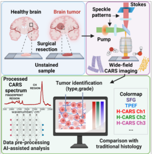
Central Nervous System (CNS) tumors encompass over 150 different types of tumors affecting the brain and spinal cord and can be categorized as primary or metastatic. Precise and rapid classification of these tumors is crucial to guide surgical decisions and plan personalized treatment strategies. Although modern imaging techniques can determine whether a CNS tumor is primary or metastatic, surgical resection followed by intraoperative histological analysis is necessary to identify the specific tumor type. Traditional histology, relying on the use of dyes, is time-consuming, lacks chemical specificity, and fails to provide a label-free, rapid, and automated tool for CNS tumor classification.
CHIMERA addresses these limitations, providing CNS tumor research with an imaging platform for intraoperative tumor diagnosis and classification that rapidly extracts spectral, morphological, and biochemical features of tumors in a label-free way. CHIMERA will merge the sensitivity of nonlinear multiphoton techniques to endogenous biomarkers with the chemical specificity of vibrational imaging approaches, using sum-frequency generation, two-photon excited fluorescence, and hyperspectral coherent anti-Stokes Raman scattering. CHIMERA will adopt a wide-field random illumination microscopy scheme that provides super-resolution in the transverse directions, z-sectioning capabilities, reduced sample damage, and unprecedented imaging speed over a large field of view. Through data processing and deep-learning classification methods, CHIMERA will offer a highly specific morpho-chemical contrast palette to simplify and accelerate the classification of various CNS tumor types. CHIMERA will advance current nonlinear microscopy technologies, unveil new insights for studying CNS tumors, and serve as a powerful diagnostic tool in biomedical research and clinical settings.
Biography : Federico Vernuccio is a CNRS post-doctoral researcher at Institut Fresnel, Marseille. After graduating with top honors from Galileo Galilei Scientific High School in Modica, Sicily, in 2014, he moved to Milan for university studies. In 2017, he earned a Bachelor’s degree with honors in Physics Engineering from Politecnico di Milano. In 2019, in the same institution, he completed his Master’s studies in Photonics and Nanooptics with honors, following an experimental thesis on developing innovative spectroscopy methods for coherent Raman techniques. The excitement of scientific work in the lab and a multidisciplinary project aimed at advancing these techniques for biomedical applications motivated him to pursue a Ph.D. in the same laboratory. His Ph.D. research has significantly advanced broadband coherent Raman scattering microscopy. A key breakthrough in his work lies in the development of a novel platform for broadband CARS, rapidly acquiring Raman spectra from cancer cells and tissues in just 1 millisecond. The key innovation behind this accomplishment is the application of a white light continuum in bulk media as a broadband source. The outcomes of these research works have been acknowledged and celebrated on a global scale, with 10 publications in peer-reviewed journals and acceptance for 12 oral presentations, 1 invited talk, and 1 poster at esteemed Photonics conferences worldwide. In 2024, he received the prize for the best 2023 doctoral thesis in physical disciplines on the topic “Applications of advanced optical methodologies in research, medicine, and telecommunications” by Accademia Pontaniana, University of Naples Federico II.
