Principal investigators : Serge Monneret, Julien Savatier
Keywords : lateral shearing interferometry, label-free microscopy, dry mass measurement of living cells
1) Principle of phase and retardance microscopies by wavefront analysis
Phase microscopy:
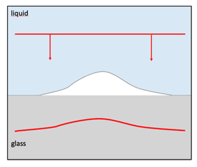 If we consider that changes in the refractive index within a transparent sample introduce local photon delays with respect to one another, sending an optical wave on this sample will result in producing a distorted wavefront as a result of propagating within such an inhomogeneous medium. Recording the shape of the resulting wavefront directly gives a spatial distribution of the so-called optical path difference OPD(x,y) defined by:
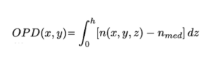
Such a distribution provides us with a complete understanding of the constitution of the object. The technique we propose here consists of simply measuring the wavefront after the sample perturbation, using a wavefront sensor, and then deducing the optical thickness mapping of this sample knowing beforehand the shape of the incident wavefront. This technique has been made possible by the emergence of wavefront sensors with sufficient resolution to produce useful information.
In this case, all of the work presented here was carried out using wavefront analyzers based on an interferometric technique called QLSI (Quadriwave Lateral Shearing Interferometry), marketed by the French company Phasics, with whom a long-term collaboration (2009-present) has been established.
Retardance microscopy:
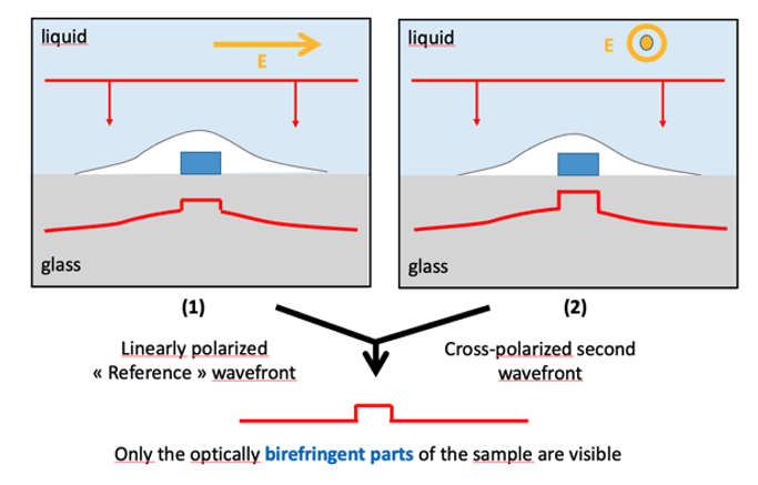 As explained before, to obtain a map of the OPD for a given sample, we successively measure two wavefronts in unpolarized light, namely a first one outside the sample (reference wavefront) and a second one with sample. A possible variation of the technique consists of using a linearly polarized incident light beam and measure a first wavefront with polarization along a defined axis, then a second with crossed polarization, perpendicular to the first one. Consequently, if the observed sample is optically isotropic, the two wavefronts will be identical. On the other hand, if some area in the sample (blue area on the figure) is optically birefringent, then the refractive index, and thus the retardance, seen by the light field, will be different for the two polarization states under consideration. A comparison between these two wavefronts then allows us to specifically identify any birefringent structure (within the observation plane) of the sample.
To find out more:
Basics of quadriwave lateral shearing interferometry for quantitative phase microscopy
P. Bon, G. Maucort, B. Wattellier, S. Monneret, “Quadriwave lateral shearing interferometry for quantitative phase microscopy of living cells”, Optics Express 17 (15), 13080-13094 (2009).
https://doi.org/10.1364/OE.17.013080
Retardance microscopy
S. Aknoun, P. Bon, J. Savatier, B. Wattellier, S. Monneret, “Quantitative Retardance Imaging of biological samples using Quadri-Wave Lateral Shearing Interferometry”, Optics Express 23 (12), 16383-16406 (2015).
https://doi.org/10.1364/OE.23.016383
Book chapter
S. Monneret, J. Savatier, P. Bon, « Quantitative Phase Microscopy Using Wavefront Analysis », in Unconventional Optical Imaging for Biology, John Wiley & Sons, ISBN: 978-1-394-28398-9 (2024).
https://doi.org/10.1002/9781394283996.ch1
Phase reconstruction
Bon, S. Monneret, B. Wattellier, « Non-iterative boundary-artifact free wavefront reconstruction from its derivatives », Applied Optics 51 (23), 5698-5704 (2012).
https://doi.org/10.1364/AO.51.005698
Gouy phase shift imaging
P. Bon, B. Rolly, N. Bonod, J. Wenger, B. Stout, S. Monneret, H. Rigneault, “Imaging the Gouy phase shift in photonic jets with a wavefront sensor”, Optics Letters 37 (17), 3531-3533 (2012).
https://doi.org/10.1364/OL.37.003531
Vibrational phase imaging
P. Berto, D. Gachet, P. Bon, S. Monneret, H. Rigneault, « Wide-field vibrational phase imaging », Phys. Rev. Lett. 109, 093902 (2012).
https://doi.org/10.1103/PhysRevLett.109.093902
P. Berto, A. Jesacher, C. Roider, S. Monneret, H. Rigneault, M. Ritsch-Marte, “Wide-field vibrational phase imaging in an extremely folded box-coherent anti-Stokes Raman scattering geometry”, Optics Letters 38 (5), 709-711 (2013).
https://doi.org/10.1364/OL.38.000709
Tomographic imaging
Y. Ruan, P. Bon, E. Mudry, G. Maire, P. Chaumet, H. Giovannini, K. Belkebir, A. Talneau, B. Wattellier, S. Monneret, A. Sentenac, “Tomographic diffractive microscopy with a wavefront sensor”, Optics Letters 37 (10), 1631-1633 (2012).
https://doi.org/10.1364/OL.37.001631
3D localization of metallic nanoparticles
P. Bon, N. Bourg, S. Lécart, S. Monneret, E. Fort, J. Wenger, S. Lévêque-Fort, “Three dimensional nanometer localization of nanoparticles to stabilize super-resolution microscopy”, Nature Comm. 6, 7764 (2015).
https://www.nature.com/articles/ncomms8764
Tomographic imaging
Bon, B. Wattellier, S. Monneret, « Modeling quantitative phase image formation under tilted illuminations », Optics Letters 37 (10), 1718-1720 (2012).
https://doi.org/10.1364/OL.37.001718
3D imaging
P. Bon, S. Aknoun, S. Monneret, B. Wattellier, “Enhanced 3D spatial resolution in quantitative phase microscopy using spatially incoherent illumination”, Optics Express 22 (7), 8654-8671 (2014).
https://doi.org/10.1364/OE.22.008654
High-definition phase imaging
Benoit Wattelier, Anais Saintoyant, Julien Savatier, Lucie De Laulanié, Sherazade Aknoun, Roman Zinchuk, S. Monneret, “High-definition quadriwave lateral shearing interferometry”, JOSA A 41 (11), C99-C108 (2024).
https://doi.org/10.1364/JOSAA.533811
2) Phase microscopy used as a measurement tool
The technique we have developed makes it possible to measure the deformation of a wavefront after it has passed through a sample of interest. As a result, we obtain the OPD map of the sample. Once we are able to decorrelate local thickness and refractive index, these OPD values allow us to characterize certain parameters of the sample, such as its topography (if the index is known) or its index (if the shape is known). More generally, we were able to carry out such measurements by changing the environmental conditions of the sample (index based on temperature, concentration, pressure, etc.).
To find out more:
Optical profilometry on etched surfaces, laser damage craters
D.-B. Douti, M. Chrayteh, S. Aknoun, T. Doualle, C. Hecquet, S. Monneret, L. Gallais, “Quantitative phase imaging applied to laser damage detection and analysis”, Applied Optics 28 (54), 8375-8382 (2015).
https://doi.org/10.1364/AO.54.008375
T. Doualle, L. Gallais, S. Monneret, S. Bouillet, A. Bourgeade, C. Ameil, L. Lamaignère, P. Cormont, “CO2 laser microprocessing for laser damage growth mitigation of fused silica optics”, Optical Engineering 56 (01), 011022 (2017).
https://doi.org/10.1117/1.OE.56.1.011022
T. Doualle, A. Ollé, S. Monneret, L. Gallais, “Laser-induced birefringence measurements by quantitative polarized phase microscopy”, Optics Letters 42 (8), 1616-1619 (2017) ;
https://doi.org/10.1364/OL.42.001616
Characterization of optical properties of materials
S. Khadir, P. Bon, D. Vignaud, E. Galopin, N. McEvoy, D. McCloskey, S. Monneret, G. Baffou, “Optical imaging and characterization of graphene and other 2D materials using quantitative phase microscopy”, ACS Photonics 4(12), 3130-3139 (2017).
https://doi.org/10.1021/acsphotonics.7b00845
Viola V. Vogler-Neuling, Romolo Savo, David Pohl, Nicholas R. Hendricks, Lukas Lang, Maria Timofeeva, Barbara Schneider, Felix Richter, Flavia Timpu, Serge Monneret, Fabian Starsich, and Rachel Grange, “Solution-Processed Barium Titanate Nonlinear Woodpile Photonic Structures with Large Surface Areas”, Physica Status Solidi B, 1900755 (2020).
https://doi.org/10.1002/pssb.201900755
J. Herbuvaux, F. Wagner, L. Vazquez-Zuniga, S. Monneret, J. Nillon, C. Hönninger, L. Gallais, “Characterization and simulation of laser-induced contamination effects on laser beam propagation in the UV sub-picosecond regime”, Applied Optics 64(17):4922-4933 (2025).
https://doi.org/10.1364/AO.559241
N. Jebali, C. Molinaro, J. Roul, J. Jermann, J.-B. Doucet, B. Reig, S. Monneret, O. Haeberlé, O. Soppera, V. Bardinal, “Self-written high-efficiency single-mode optical link using a single near-infrared photopolymerization step”, J. of Lightwave Techn. PP(99):1-7 (2025).
https://doi.org/10.1109/JLT.2025.3628991
Laser-induced effects measurements
L. Gallais, S. Monneret, “Time-resolved quantitative phase microscopy of laser-material interactions using a wavefront sensor”, Optics Letters 41(14), 3245-3248 (2016).
https://doi.org/10.1364/OL.41.003245
M. Soulier, A. Rapaport, H. Krol, S. Monneret, J. Lumeau, L. Gallais, “Measurement and simulation of wavefront deformation induced by a high-power CW laser in thin-film optical filters”, Applied Optics 65(5):A68-A78 (2025)
https://doi.org/10.1364/AO.575022
Thermo-refractive index (dn/dT) measurement of liquids
R. Radhakrishnan, L. Gallais, S. Monneret, “Wavefront sensing applied to determine temperature dependence of liquids”, Applied Optics 58(13), 3646-3651 (2019).
https://doi.org/10.1364/AO.58.003646
Temperature microscopy
G. Baffou, P. Bon, J. Savatier, J. Polleux, M. Zhu, M. Merlin, H. Rigneault, S. Monneret, « Thermal imaging of nanostructures by quantitative optical phase analysis », ACS Nano 6 (3), 2452-2458 (2012).
https://doi.org/10.1021/nn2047586
3) Phase microscopy used as an added value for biology
The initial motivation behind our developments was to provide a highly contrasted imaging technique for living biological cells that did not require labeling, enabling biologists to visualize the condition of their samples without using fluorescence and therefore without any time limitations (no photobleaching) or modification of the gene expression (no transfection) nor direct tagging with big fluorescent molecules. However, the quantitative aspect of the measurements quickly opened up this technology to applications of great interest, such as measuring the dry mass of living biological cells, enabling the cell cycle to be tracked, or monitoring the evolution of a culture under different environmental conditions.
To find out more:
Label-free imaging / simultaneous phase-fluorescence imaging
P. Bon, J. Savatier, M. Merlin, B. Wattellier, S. Monneret, “Optical detection and measurement of living cell morphometric features with single-shot quantitative phase microscopy”, J. Biomed. Opt. 17 (7), 076004 (2012).
https://doi.org/10.1117/1.JBO.17.7.076004
Dry mass determination of cells
S. Aknoun, J. Savatier, P. Bon, F. Galland, L. Abdeladim, B. Wattellier, S. Monneret, “Living cells dry mass measurements using Quantitative Phase Imaging with Quadri Wave Lateral Shearing Interferometry. An accuracy and sensitivity discussion”, Journal of Biomedical Optics 20(12), 126009 (2015).
https://doi.org/10.1117/1.JBO.20.12.126009
C. Allier, L. Hervé, O. Mandula, P. Blandin, Y. Usson, J. Savatier, S. Monneret, S. Morales, “Quantitative phase imaging of adherent mammalian cells: a comparative study”, Biomedical Optics Express 10 (6), 2768-2783 (2019).
https://doi.org/10.1364/BOE.10.002768
Detection of collagen fibers
Aknoun, M. Aurrand-Lions, B. Wattellier, S. Monneret, “Quantitative Retardance Imaging by means of Quadri-wave lateral shearing interferometry for label-free fiber imaging in tissues”, Optics Communications 422, 17-27 (2018).
https://doi.org/10.1016/j.optcom.2018.02.061
Photothermal control of single cells
Robert, J. Savatier, S. Vial, J. Verghere, B. Wattellier, H. Rigneault, S. Monneret, J. Polleux, G. Baffou, “Photothermal control of heat-shock protein expression at the single cell level”, Small 2018, 1801910 (2018).
