Principal investigator : Thomas Chaigne
Photoacoustic tomography is a biomedical imaging technique that uses laser-induced ultrasound waves to probe optical absorption deep (> 1 mm) inside scattering biological tissue.
To generate an image, pulsed light illuminates the tissue sample. Embedded dark structures, such as blood vessels, will absorb energy and heat up briefly. This transient temperature rise is transformed into a pressure wave through thermo-elastic effect. The ultrasound wave can then propagate through soft tissue with minimal scattering, and can therefore be detected at the tissue surface.
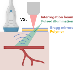
Figure 1: Photoacoustic imaging provides optical absorption contrast at large depth (>1mm) in highly scattering tissue. Optical detection of ultrasound provides larger bandwidth and increased sensitivity at high frequencies, as compared to piezoelectric sensors.
We develop optical sensors of ultrasound based on Fabry-Perot cavities with a polymer sensing layer. These sensors exhibit a broad bandwidth than conventional piezoelectric probes, offering a higher spatial resolution (down to 10 µm). We design and fabricate them in collaboration with the RCMO group and the Micro-electronics center of Provence.
So we not only generate sound with light, but we also measure sound with light. How does it work? Thanks to optical interferences in the resonant optical sensor, the reflectivity exhibits some minima. As the positions of these minima are linked to the sensor thickness, they will be slightly shifted when ultrasound waves propagate through the sensor.
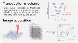
Figure 2: (top) How to measure ultrasound with light, (bottom) Acquisition scheme: an interrogation beam scans the sensor surface, and a fast photodetector records the modulated reflected intensity at each point. A 3D image is reconstructed from the measured 2D acoustic field over time.
Since our sensors are ultrathin to maximize detection bandwidth, they are highly sensitive to thickness variations in the polymer layer introduced during fabrication. A conventional system compensates for this by tuning a narrowband laser source at each measurement point, but this approach is slow and limits temporal resolution. But this turns out to be very time consuming and therefore strongly limits the temporal resolution. To address this, we implemented a fast optical interrogation strategy to compensates for fabrication tolerances while maintaining a high frame rate [Saucourt 2023].
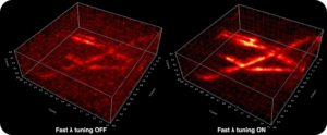
Figure 3: Photoacoustic images of 20 µm nylon wires, with (right) and without (left) compensation for thickness inhomogeneities.
To further increase the temporal resolution, we design new compressive image acquisition and reconstruction strategies in collaboration with the Comix group. These leverage the spatio-temporal properties of the targeted objects to reduce the amount of measurements required.
On top of instrumentation developments, we apply photoacoustic imaging for biology, with a particular focus on neuronal activity imaging. We collaborate closely with local neurobiology groups at Inmed and INT.
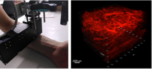
Figure 4: (left) Experimental system applied to a human subject, (right) Photoacoustic image of blood vessels (and sub-cutaneous hair) in human forearm (The PI’s arm!).
