Smart 3D fluorescence microscopy
Principal investigators : Loic Le Goff, Hervé Rigneault
Collaboration: F. Galland (Phyti group), A. Sentenac (SEMO group)
Keywords : Smart scanning, Random illumination microscopy, speckle illumination, nonlinear imaging, event cameras
3D imaging of biological tissues relies on the sequential acquisition of a large images (several Gb) over numerous optical planes to reconstruct complex volumes. However, this approach presents several major challenges:
– Limited number of points with relevant information, due the often sparse nature of the sample
– Limited imaging speed, due to the need to sequentially acquire numerous planes.
– Increased light dose, leading to phototoxicity and photobleaching.
– Significant fluorescence background, resulting in loss of contrast and potentially resolution.
– Optical aberrations induced by the tissue.
We are exploring smart imaging strategies coupled to computational imaging and novel technologies such as event cameras to address these challenges.
Smart-Scan: Reducing Light Exposure and Speeding Up Imaging
Smart-scan uses an iterative, unsupervised feedback loop between sparse acquisitions and signal analysis to pinpoint structures of interest within the tissue. Once these structures are identified, illumination is selectively targeted around them. This strategy dramatically reduces light exposure and has the potential to significantly accelerate tissue imaging.
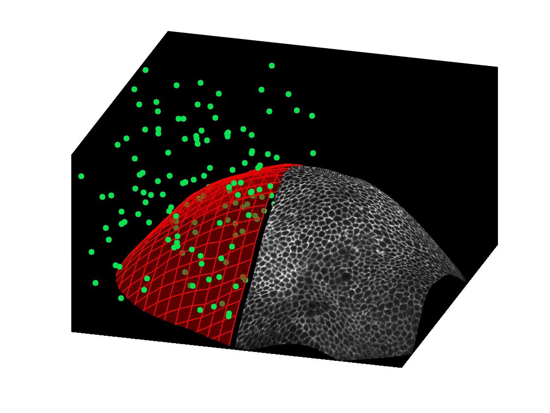
Starting from just a few acquisitions (green points), the Smart-scan microscope automatically estimates the tissue structure (red mesh) and can then focus subsequent acquisitions on those regions. This targeted approach optimizes imaging efficiency and precision.
Random Illumination Microscope (RIM) is a specialized form of structured illumination microscopy, designed specifically for complex biological environments. It leverages speckle patterns as structured illumination. This approach offers two key advantages:
– The statistical properties of speckles are inherently robust against optical aberrations.
– The RIM reconstruction process itself relies on statistical analysis (variance) of images to produce optical sectionning and ultra-resolution (x2)
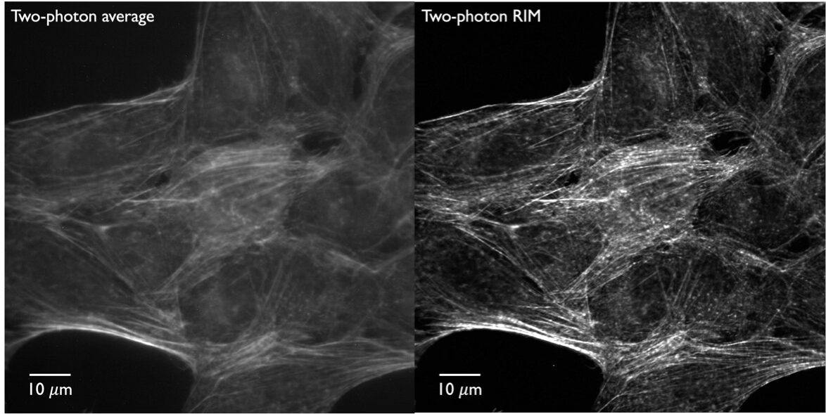
2Photon fluorescent images: Wide-field 2Photon image (left), 2Photon RIM image (optically sectionned and super-resolved) (right)
We are developping:
– Extended depth of field RIM, that combines speckle illumination, extended depth-of-field detection, and RIM reconstruction, this approach delivers super-resolved projective images in a single acquisition.
– Two-Photon RIM, provides superior penetration depths, wide-field optical sectioning and super-resolution in scattering sample.
– 3D-RIM extends the principles of RIM to fully three-dimensional acquisition and reconstruction. It leverages the inherently 3D nature of speckles and volumetric imaging through remote focusing, without the need to physically shift the illumination. This approach is highly photon-efficient and demonstrates exceptional capability for deep imaging, even in aberrated conditions.
– RIM with event-based camera and liquid crystal speckle generator: Using an event-based camera and a fluctuating random speckle illumination based on a smart liquid crystal, we perform optical sectioning wide-field fluorescence imaging in thick samples. This hardware-based approach drastically reduces the computation time and data storage requirements for optically sectioned wide-field microscopy.
– Temporal focusing with speckle illumination: To perform z-sectioning deep tissue imaging and improve RIM sectioning capability.
General audience
Smart Illumination for 3D-imaging of biological tissues. LeGoff, Mazzella, Cecchini, Allain, Sentenac, Galland (2025) Photoniques 134, p. 36. DOI: 10.1051/photon/202513436
REFERENCES:
2026
Speckle illumination temporal focusing two-photon excited fluorescence microscopy. Federico Vernuccio, Michał Marynowski, Xiangyi Li, Assia Benachir, Luca Genchi, Randy Bartels, Sandro Heuke, Anne Sentenac, and Hervé Rigneault, Optica 13, 577-586 (2026), https://doi.org/10.1364/OPTICA.581520
Optical sectioning in wide-field two-photon microscopy using temporal focusing and random illumination. Xiangyi Li, Federico Vernuccio, Michal Marynowski, Assia Benachir, Sandro Heuke, Hervé Rigneault, and Anne Sentenac, Opt. Lett. 51, 1428-1431 (2026)
https://doi.org/10.1364/OL.592916
Tunable Dynamic Speckle Generation for Random Illumination Microscopy, Advanced Materials Technologies, L. Magermans, A. Benachir, N. P. Spiller, T. Wang, F. Vernuccio, R. Bartels, S. M. Morris, S. J. Elston, M. J. Booth, and H. Rigneault, on line publication e02098. DOI: https://doi.org/10.1002/admt.202502098
2025
Smart Illumination for 3D-imaging of biological tissues. LeGoff, Mazzella, Cecchini, Allain, Sentenac, Galland (2025) Photoniques 134, p. 36. DOI: 10.1051/photon/202513436
Super-resolved live imaging of thick biological samples with 3D Random Illumination Microscopy (3D-RIM).Mangeat, Mazzella, Rogez, Giroussens, Bernard, Vargas, Allain, Labouesse, Idier, LeGoff, Sentenac (2025). BioRxiv. DOI: 10.1101/2025.08.06.668879
2024
Fast super-resolved reconstructions in fluorescence random illumination microscopy (RIM). Giroussens, Labouesse, Allain, Mangeat, Mazzella, LeGoff, Sentenac, Idier, IEEE Transactions on Computational Imaging. (2024) DOI: 10.1109/TCI.2024.3507643
Extended-depth of field random illumination microscopy, EDF-RIM, provides super-resolved projective imaging. Mazzella, Mangeat, Giroussens, Rogez, Li, Creff, Saadaoui, Martins, Labouesse, Idier, Galland, Allain, Sentenac, LeGoff, (2024) Light, science and applications DOI: 10.1038/s41377-024-01612-0.
2023
Adaptive scans allow 3D-targeted laser-dissection to probe the mechanics of cell sheets. Meng, Nuzdhin, Sison, Galland, LeGoff (2023) EPJ Plus, https://rdcu.be/dj9Ms DOI : 10.1140/epjp/s13360-023-04378-3
2022
Optimizing sampling for surface localization in 3D-scanning microscopy, M.-A. Burcklen, F. Galland, and L. Le Goff, Journal of the Optical Society of America A 39, 1479-1488 (2022). https://doi.org/10.1364/JOSAA.460077
2021
An adaptive microscope for the imaging of biological surfaces,F. Abouakil, H. Meng, M.-A. Burcklen, H. Rigneault, F. Galland, and L. LeGoff, Light: Science & Applications 10, 210 (2021).
https://doi.org/10.1038/s41377-021-00649-9
