Polarized fluorescence microscopy
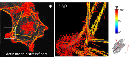
Light polarization can be advantageously coupled to fluorescent molecules to probe their orientations. Using excitation polarization tuning, it is possible to measure the orientational order of fluorescent molecules attached to biological proteins or lipids of interest, within the focal volume of excitation. We have developed methods based on confocal microscopy, spinning disk imaging and two- and three-photon imaging, sensitive to the excitation polarization.
Highlights:
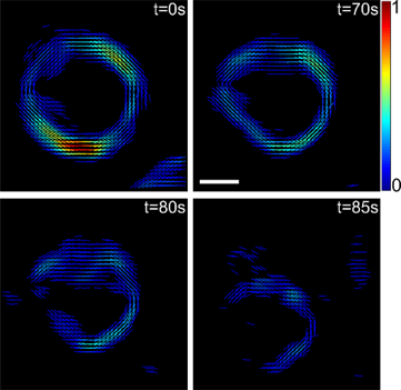
- Molecular order imaging in actin in the cell cytoskeleton
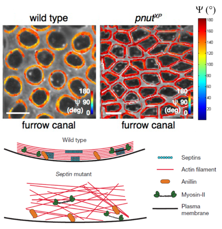
- Molecular order imaging in actin in the cytokinesic ring of cells during cellularization in the drosophila melanogaster embryo.
M. Mavrakis, Y. Azou-Gros, F-C. Tsai, J. Alvarado, A. Bertin, F. Iv, A. Kress, S. Brasselet, G.H. Koenderink and T. Lecuit.
Septins promote F-actin ring formation by cross-linking actin filaments into curved bundles
Nature Cell Biology 16, 322–334 (2014) - Molecular order imaging in actin in the cytokinesic ring of cells during cellularization in the drosophila melanogaster embryo.
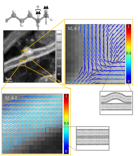
A. Kress, X. Wang, H. Ranchon, J. Savatier, H. Rigneault, P. Ferrand, S. Brasselet
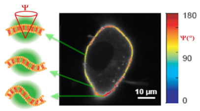
Mapping the local organization of cell membranes using generalized polarization resolved confocal fluorescence microscopy
Biophys. J. 105, 127-136 (2013)
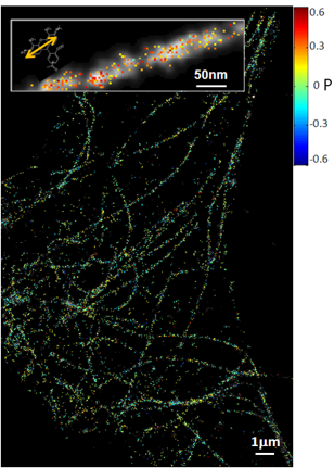
Polarized super-resolution imaging
Fluorescence polarized microscopy is scaled down to the single molecule scale, in order to (1) get more information on the orientational flexibility of the fluorophores that are linked to proteins, (2) reconstruct super-resolution orientation images using their localization information. Orientational flexibility is an important parameter to quantify to be able to retrieve realistic structural information on the labeled bio-molecules. A wide field polarized fluorescence experiment is developed that can give both mean orientation and wobbling extent for each single molecule attached to filament structures in vitro and in cells. This approach is applied to organized filaments such as dsDNA, actin stress fibers, microtubules, insulin amyloids, and is currently extended to membrane proteins. Super-resolution orientation images are reconstructed using polarized dSTORM (direct Stochastic Optical Reconstruction Microscopy) and PALM (photoactivated light microscopy).
Highlights:
- Imaging of fluorophores orientation and fluctuations in filaments of the cell cytoskeleton
C.A. Valades Cruz, H. A. Shaban, A. Kress, N. Bertaux, S. Monneret, M. Mavrakis, J. Savatier, S. Brasselet
Quantitative nanoscale imaging of orientational order in biological filaments by polarized super-resolution microscopy
Proc. Natl. Acad. Sci. 113, (7) E820-E828 (2016)
Nonlinear microscopy
Due to the nonlinear vectorial coupling between incident light polarizations and molecular bonds / molecular induced dipoles directions, tunable incident polarizations lead to a modulation of nonlinear label-free signals (Second Harmonic Generation : SHG, Coherent Anti Stokes Raman Scattering : CARS, Stimulated Raman Scattering : SRS) that can be directly related to the orientational order of biological molecules, without the need of a fluorescent label. We have applied polarized nonlinear microscopy to quantitatively retrieve organizational order in collagen in tissues (by polarized SHG) and lipid structures (by polarized CARS sensitive to CH2 stretching vibrations). This methodology is highly sensitive to lipid phases but also to sub-diffraction scale morphological changes in cell membranes. Polarization resolved CARS (pCARS) is now used to image the effect of neurodegenerative diseases on fine myelin structural changes, in mice spinal cord tissues.
Highlights:
- Molecular order imaging of lipids in the myelin sheath of the mouse spinal cord axons using label free, chemically sensitive coherent anti Stokes Raman scattering
P. Gasecka, A. Jaouen, F.-Z. Bioud, H. Barbosa de Aguiar, J. Duboisset, P. Ferrand, H. Rigneault, N. Balla, F. Debarbieux, S. Brasselet
Degradation of molecular organization of myelin lipids in autoimmune demyelination probed by polarization resolved nonlinear vibrational microscopy
Biophys. J. accepted (2017)
 |
M. Hofer, N.K. Balla, S. Brasselet |
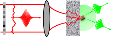
Imaging in scattering media
We have recently shown that the use of broadband wavefront shaping to refocus light through scattering media permits to conserve time and polarization properties so as to performe nonlinear optical imaging (second harmonic generation) and polarized imaging.
Highlights:
- Nonlinear imaging through a scattering medium.
H. B. De Aguiar, S. Gigan, S. Brasselet
Enhanced nonlinear imaging through scattering media using transmission matrix based wavefront shaping
Phys. Rev. A 94, 043830 (2016) - Polarized recovery by broadband wavefront shaping through a scattering medium
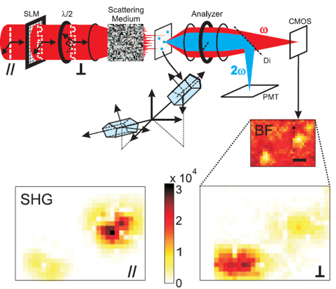
H.B. De Aguiar, S. Gigan, S. Brasselet
Polarization recovery through scattering media
Sci. Adv. accepted (2017), arXiv:1511.02347
Nanophotonics
We have demonstrated structural analysis of metal nano-structures supporting local enhancement of optical responses, as well as the diagnostics of mono-crystallinity in inorganic nanocrystals. This properties takes advantage of the polarization dependence of second harmonic generation to nanoparticules surface and symmetry, to reveal sub-diffraction scale features related to their shape or crystalline phases.
Highlights:
- Vectorial imaging of nonlinear fields enhancements in plasmonics nanoparticles, at the nanometric scale
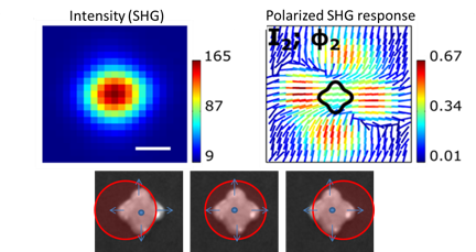
N.K. Balla, C. Rendon-Barraza, L.M. Hoang, P. Karpinski, E. Bermudez, S. Brasselet
Polarized nonlinear nanoscopy of metal nanostructures
ACS Photonics accepted (2017), 10.1021/acsphotonics.6b00635
Funding