
TexTOM : La «texture tomography» par rayons X comme outil permettant l’imagerie multi-échelle et in-situ de l’enthèse, connexion biologique entre l’os et le tendon
Début du projet : 01.07.2022
Fin du projet : 30.6.2027
Résumé du Projet
Biological materials exhibit outstanding mechanical properties and combine seemingly contradictory features like strength and toughness. This is caused by the intricate, hierarchical organization of these materials on the nanostructural and crystal structural level, the so-called crystallographic texture. The determination of the crystallographic texture and the local nanostructure in 3D, while retaining a large field of view is an unsolved problem until now.
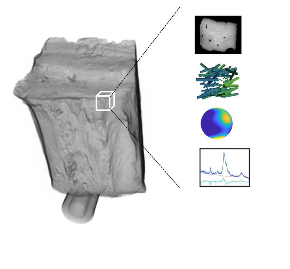
In the framework of the ERC Starting grant project “TexTOM”, this problem is going to be solved by introducing texture tomography, a 3D imaging technique based on X-ray diffraction and employed a synchrotron radiation source, schematically sketched in Fig 1.
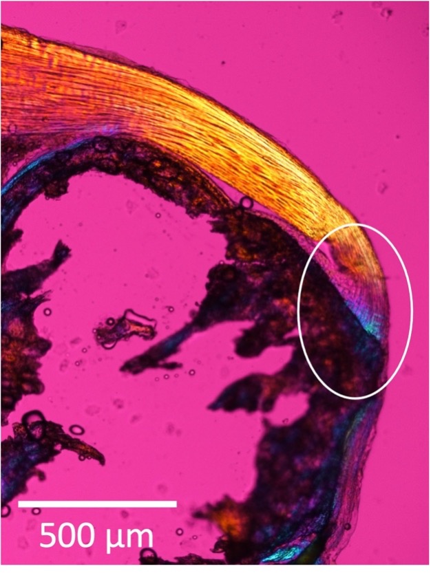
With this technique, it will be possible to understand the structure of the enthesis (Fig 2), the biological connection between bone and tendon, which is often involved in orthopedic injuries.
Together with his collaborators at the Institute des Sciences du Mouvement, Marseille, he will establish the relation between the hierarchical organization and the mechanical properties of the enthesis and use this information to develop a micromechanical model of the enthesis.
Mots Clés : Imagerie Rayon X, Diffraction, Textures Crystallographiques, Biominéralisation, Biomécanique
Partenaires
Institut des Sciences du Mouvement, Marseille
European Synchrotron Radiation Facility, Grenoble, France
Aarhus University, Aarhus, Denmark
Paul Scherrer Institute, Villigen, Switzerland
Vienna University of Technology, Vienna, Austria
Biographie
Tilman Grünewald is a CNRS researcher in the COMiX team at the Institut Fresnel. His research aims at understanding the structure and behavior of biomineralized tissues with the help of X-ray scattering approaches, in particular by developing means to study the 3D arrangement of the crystalline building blocks and the nanostructure of materials.
In the past years, he has focused his research activity on the development of new approaches and uses them to understand the structure of biological tissues. Examples are the structure bone and its interplay with bioresorbable implants or the development of the crystalline structures of oyster shells. To this end, he develops combined in-vivo and in-situ experiments couple with synchrotron x-ray diffraction approaches. For this, he is working jointly with synchrotron beamlines to expand the experimental portfolio as well as collaborating with experts in the field of biomineralization.
His research program has been recently awarded with an ERC Starting grant which will help him to develop texture tomography as a tool for quantitative texture information in 3D and use it to determine the structure and load transfer properties of the enthesis, the biological connection between tendon and bones.
 ResearchGate
 Flux RSS