
Contact: Anabela Da Silva
Photoacoustic tomography (PAT) is a multiwave imaging modality that potentially combines the high optical contrast brought by the optics and high spatial resolution by ultrasound probing. The interests in using this technique for biomedical sensing are growing since 1990s, and its applications are in breast cancer detection, small animal molecular imaging, subcutaneous structures imaging, functional imaging, and other applications.
The principle consists in illuminating a sample with a local absorbing optical contrast (vasculature, presence of a tumor for example). The absorbed energy converts into heat and the temperature of the object increases, causing thermal expansion that generates an acoustic pressure wave. The energy deposited is proportional to the fluence and the local absorption coefficient of the tissues. Hence, the primary aim of PAT is to retrieve the spatial 3D distribution of the absorption coefficient of the probed tissue. This absorption coefficient, proportional to the concentration of the different chromophores composing the tissue, is an intrinsic marker of the physiology and the metabolism. In the red and near infrared, PAT has been applied to mapping hemoglobin concentrations that can reveal the presence of a cancer, through oxy- and deoxy-hemoglobin concentrations which are markers of the oxygen consumption in the tissues. Glucose monitoring with PAT has also been reported. However, usually the technique is applied to probing tissues at small depths (small animals, subcutaneous examination), for which the fluence can be modelled as a Beer-Lambert’s law by considering light scattering is not perturbing too much the quantification. For deep tissues imaging, because of their high level of scattering, diffusion of light has to be taken into account in the evaluation of the fluence that depends non-linearly on both the absorption and diffusion coefficients. Isolating the absorption contribution from the energy deposited is the challenge in quantitative PAT (QPAT), as the fluence does depend itself on the optical parameters (absorption and diffusion coefficients) distributions.
Considering sources and detectors distributions for quantitative photoacoustic tomography
There exist basically four approaches in QPAT which consists in recovering:
i. the absorption coefficient directly. Supposing the diffusion coefficient is known;
ii. the absorption coefficient and the fluence. The fluence contribution can be filtered by making use of a contrast agent of known absorption coefficient with photoacoustic signal measurements performed before and after injection. Acousto-optics or Diffuse Optical Tomography (DOT) are also methods allowin explicitly to measure the fluence or the optical parameters. Another method consists also to introduce prior knowledge on the spatial frequency of the two quantities, high frequency for the absorption coefficient and low frequency for the fluence .
iii. the absorption and diffusion coefficients simultaneously by introducing prior knowledge (spectra of known species, diffusion spectral profile) with multiple wavelength illuminations.
iv. the absorption and diffusion coefficients simultaneously by introducing active probing with multiple optical illuminations, that is with structured illumination (multiple point-like sources for example).
With this last approach, the probing becomes active and similar with what occurs in geophysics when searching for buried objects, petroleum layers, cracks... We have adopted this approach and tested it under different sources and detectors geometries in order to test its robustness under potential experimental situations with single wavelength illumination, point scanning single element transducer and limited view angle examination [1].
Through a simulation study, it has been demonstrated that using wide field multiple illuminations improves the reconstructions (Fig. 1).
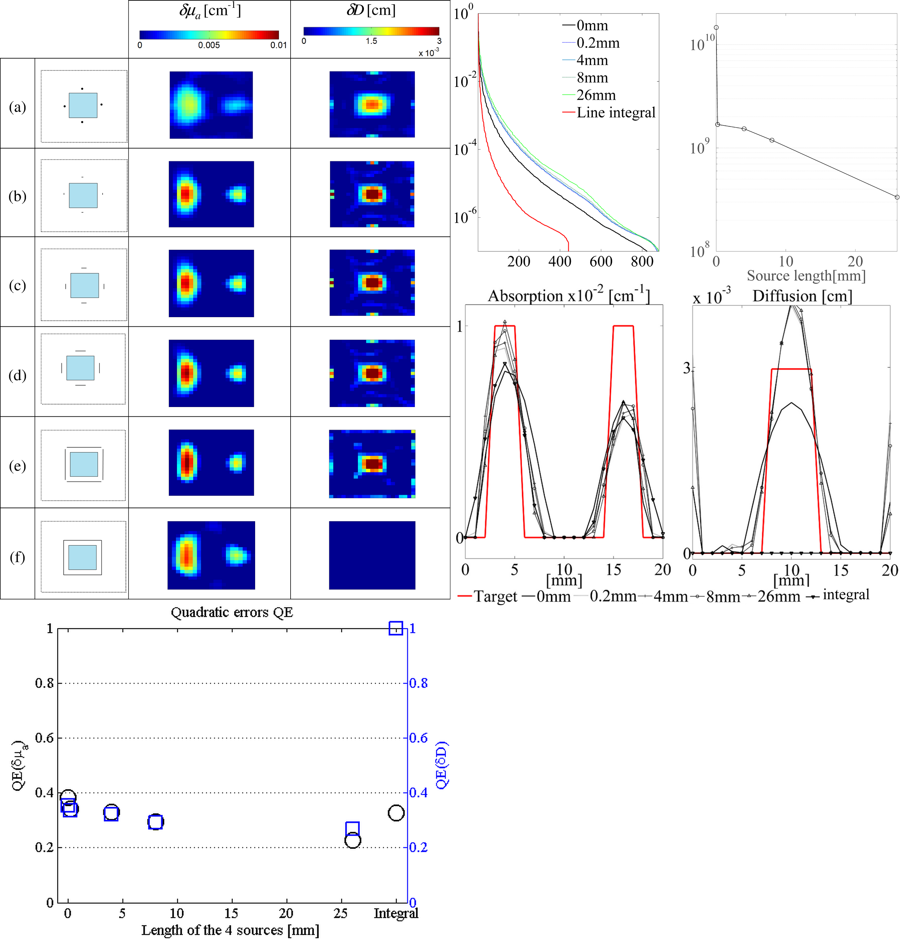
Figure 1:Influence of the detectors distributions. Left Top: reconstructed perturbation absorption and diffusion coefficient maps. (a) the detectors are evenly distributed around the object (15 detectors on each side); (b) on each side, the detectors cover a length that is half the length of the object; (c) the length covered by the detectors is half of (b); (d) 30 detectors evenly distributed on each of the two vertical sides of the object; (e) 30 detectors evenly distributed on each of the two horizontal sides of the object. Left Bottom: Quadratic errors on the reconstructions of the absorption (black circles) and diffusion (blue squares) coefficients perturbations for the five different configurations. Right Top: Normalized singular values (Left) and condition number of H as a function of the number of sources (Right). Right Bottom: cross-plots of the values extracted from a horizontal line in the middle of the vertical axis in the reconstruction areas.
Taking advantage of acoustic inhomogeneities in photoacoustic measurements
Another advantage, not exploited so far, of performing local illumination is that it generates a photoacoustic wave near the interface (Fig. 2).
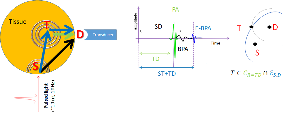
Figure 2: Schema illustrating the principle: left, a sample containing an optical and acoustic abnormality centered at position T is locally illuminated at the boundary in S by a pulsed laser (pulse width ∼ few ns) that generates two simultaneous photoacoustic waves; middle: three echoes can be probed with a transducer located at position D: the PA and BPA signals, and E-BPA resulting from the perturbation of the BPA by the presence of the acoutic abnormality; right: the time-delay of PA signal localizes the abnormality on a circle while the one of the E-BPA localizes it on an ellipse.
Usually, this signal is time-filtered or not measured (transducers oriented in specific directions). However, this PA wave, originating from the external boundary of the object (which we refer to as the Boundary PA (BPA) wave), generally follows a longer ray path within the object. Therefore,it is more likely to be scattered by any of the acoustical heterogeneities than PA waves. The BPA wave brings spatial information that can be introduced afterwards in the PA signal processing to retrieve the optical properties.
A protocole of measurement of these BPA signals, a signal processing algorithm exploiting these echographic signals for localization of embedded abnormalties (tumors) and a reconstruction algorithm have been developped (Fig. 3).
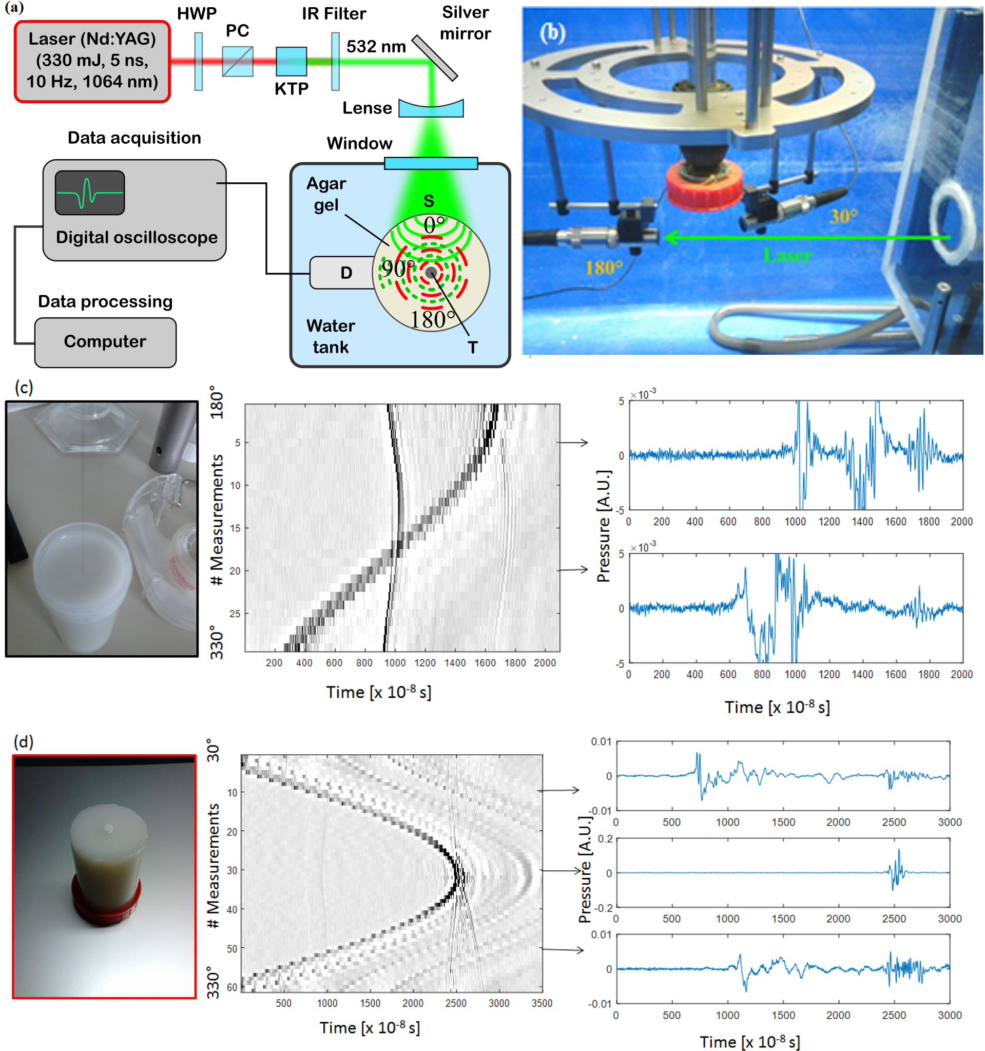
Figure 3: (a) Schema of the experimental setup;(b) photograph of the tranducers and phantom holder; (c) phantom containing hair inclusion: photograph, sinogram of the 32 measurements from 180° (transmission) to 330°,and two extracted crossplots; (d) phantom containing a purely acoustically contrasted inclusion, sinogram of the 64 measurements from 30° (reflection) to 330° (reflection), and three extracted crossplots.
Quantitative Thermoacoustic Tomography with microwaves sources- QTAT
Within ANR AVENTURES (2013-2016), in collaboration with researchers from Universities of Orléans (MAPMO lab, France) and Vienna (Computational Science Center, Austria) and from other research teams in Institut Fresnel (ILM and HIPE), a complete forward model for ThermoAcoustic Tomography (TAT) has been derived from Maxwell’s equations for the description of the deposited fluence. An inverse problem formulation is also proposed [2].
[1] Ningning Song, Carole Deumié and Anabela Da Silva, “Considering Sources and Detectors Distributions for Quantitative PhotoAcoustic Tomography,” Biomedical Optics Express 5(11), 3960-3974, 2014.
[2] H. Akhouayri, M. Bergounioux, A. Da Silva, P. Elbau, A. Litman, L. Mindrinos, “Quantitative Thermoacoustic Tomography with microwaves sources,” J. of Inverse and Ill-Posed Problems, accepted for publication novembre 2016.
This work has been supported by:
C’NANO PACA 2010
CSC Scholarship (Ningning Song)
ANR AVENTURES-ANR-12-BLANBS01-0001-04
SATT SE programme de maturation Mammoscan
 ResearchGate
 Flux RSS