
Fluorescence microscopy in the visible range is a widely used tool in biology as it is well-suited for studying living cells. Unfortunately, the resolution of this imaging technique is limited by the diffraction process, with the lateral resolution being at best 200 nm. In the SEMO team, we are developing a simple optical imaging system that offers a resolution of less than 100 nm.
Our imaging system is based on two main ideas. The first is to etch the sample holder on which the sample is deposited to create a nanostructured network. This network transforms the incident wave into evanescent waves of very high spatial frequencies that can probe the finest details of the sample.
The second idea is to illuminate this thin slice with a spatially controlled beam in amplitude and phase. Depending on the illumination mode, one can either create a light grid or continuously move a small-sized light spot on the surface of the network below the diffraction limit. This system will allow for ultra-high-resolution wide-field imaging with the light grid, and the study of dynamic phenomena through Fluorescence Correlation Spectroscopy with the spot.
This system will allow, thanks to the light grid, to do ultra-resolved wide-field imaging and, with the spot, to study dynamic phenomena by Fluorescence Correlation Spectroscopy.
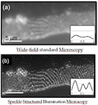
We have also shown that by illuminating the sample in a heterogeneous and random way, it is possible to improve the resolution of the microscope by a factor of two. Our approach consists in recording several images of the same sample by illuminating it with random light patterns (speckle) obtained by moving a piece of rough plastic on the path of a laser beam. An inversion algorithm is then used to reconstruct the sample from the images.
The advantage of our approach compared to other super-resolution microscopy techniques (such as confocal microscopy or structured illumination microscopy...) is that there is no need to control the illumination. This greatly simplifies the experimental implementation.
Références :
- E. Mudry, K. Belkebir, J. Girard, J. Savatier, E. Le Moal, C. Nicoletti, M. Allain and A. Sentenac, Structured illumination microscopy using unknown speckle patterns, Nature Photonics 6, 312 (2012).
- A. Sentenac, K. Belkebir, H. Giovannini and P. C. Chaumet, High resolution Total Internal Reflection Fluorescence Microscopy using periodically nanostructured glass slides, J. Opt. Soc. Am. A 26, 2550 (2009).
- A. Sentenac, K. Belkebir, H. Giovannini and P. C. Chaumet, Sub-diffraction resolution in total internal reflection fluorescence microscopy with a grating substrate, Opt. Lett. 33, 255 (2008).
- A. Sentenac, K. Belkebir, H. Giovannini, P. C. Chaumet, High resolution Total Internal Reflection Fluorescence Microscopy using periodically nanostructured glass slides, J. Opt. Soc. Am. A 26, 2550 (2009).
- A. Sentenac, K. Belkebir, H. Giovannini, and P. C. Chaumet, Sub-diffraction resolution in total internal reflection fluorescence microscopy with a grating substrate, Opt. Lett. 33, 255 (2008).
 ResearchGate
 Flux RSS