
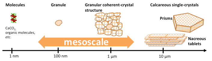
Biocrystallization involves extraordinary complex and regular biochemical processes where living organisms control the crystalline form and texture of their organo-mineral components. Although these crystals present a wide variety of shapes, associated to various organic materials, the observation of a nanoscale granular structure common to almost all calcareous crystallizing organisms, associated to an extended crystalline coherence, underlies a generic biomineralization and assembly process. A key to building realistic scenarios of biomineralization is to reveal the crystalline architecture, at the mesoscale, (i. e., over a few granules), which none of the existing nano-characterization tools is able to provide.
This study is primarily based on the exploitation of the synchrotron coherent x-ray diffraction microscopy we have developed, which gives access to the 3D mesoscale image of the crystalline properties (crystalline coherence, crystal plane tilts and strains) with the required flexibility, nanoscale resolution, and non-invasiveness. It is combined to optical ptychography (birefringency properties) and coherent anti-Stokes Raman spectroscopy (CARS).
We look at revealing the generics of the mesoscale crystalline structure of CaCO3 biominerals through the explorations of a vast variety of crystalline biominerals produced by marine calcifiers such as e. g., Pinctada margaritifera oyster shells, nacreous pearls, foraminifera, coccolithophores, etc…
Fundings: This work is funded by the French ANR under contract no ANR-11-BS10-0005 and by the European Research Council (ERC) under the European Union’s Horizon H2020 research and innovation program grant agreement No 724881.
References
 ResearchGate
 Flux RSS