3D-RIM
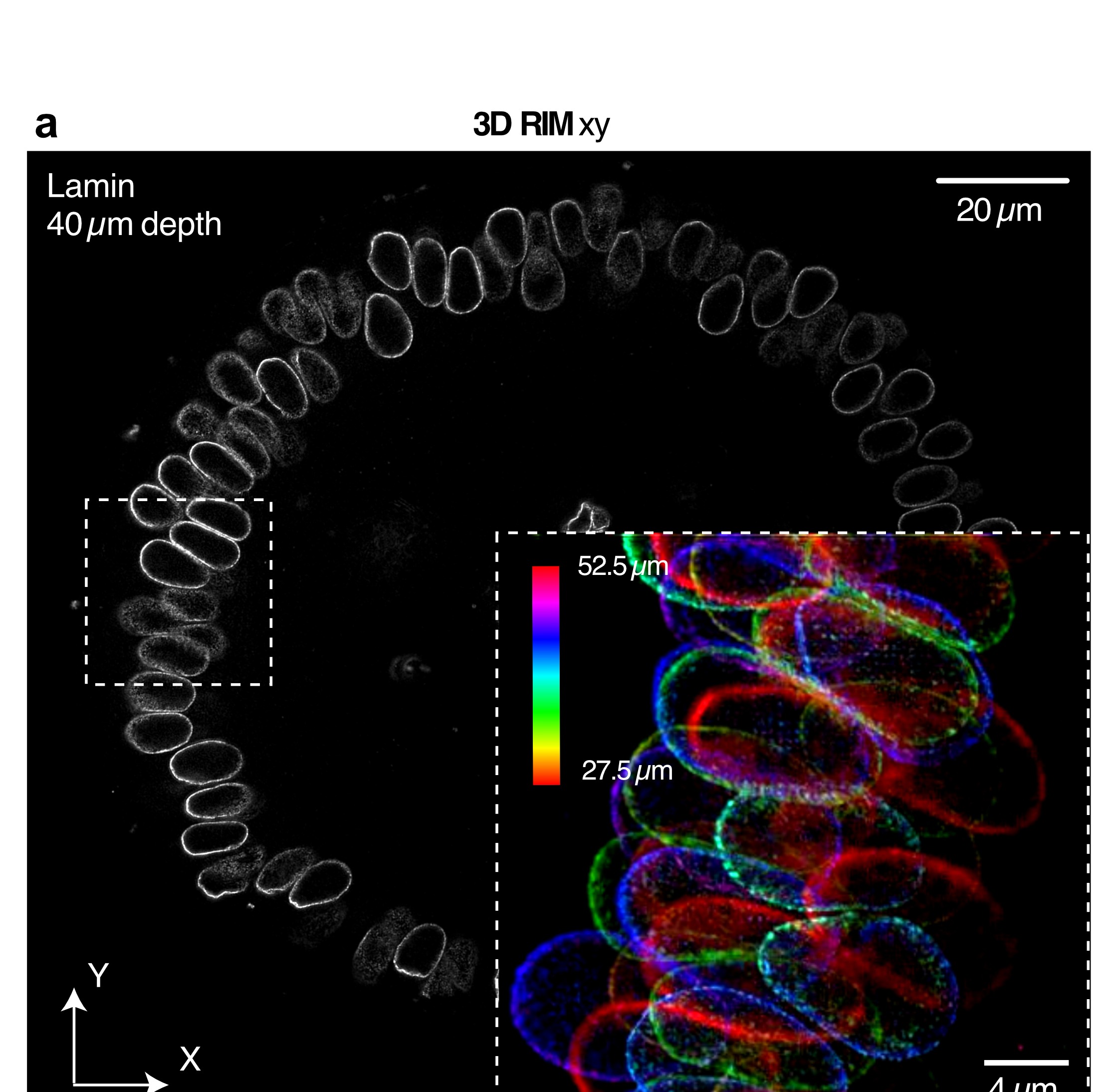
Super-resolved volume imaging of thick, live samples is hard: aberrations blur patterned illumination, and out-of-focus fluorescence kills contrast. 3D-RIM extends random illumination microscopy to full volumes using speckled illuminations, remote focusing (so the sample stays fixed relative to the illumination), 3D photon reassignment, and variance processing.
On thin samples we reach about 110 nm laterally and 270 nm axially, matching 3D-SIM. In highly aberrant tissues the method keeps high contrast over tens of microns (for example Drosophila embryos and mouse intestine), and still works hundreds of microns deep in collagen scaffolds. Live border-cell migration in the egg chamber can be followed for tens of minutes without obvious bleaching or phototoxicity.
Publications
Mangeat, Mazzella, Rogez, Giroussens, Bernard, Vargas, Allain, Labouesse, Idier, LeGoff*, Sentenac* (2025). Super-resolved live imaging of thick biological samples with 3D Random Illumination Microscopy (3D-RIM). BioRxiv. link